


Project Details
- Category Computer Vision and Digital Ophthalmology
- Date June 21, 2026
- rating

Retinal Disease Classification
Optical Coherence Tomography (OCT) is a vital, non-invasive imaging technology that provides high-resolution cross-sectional views of retinal structures. However, interpreting OCT images requires specialized expertise and considerable time.
To address this challenge, we developed a specialized Convolutional Neural Network (CNN) model capable of analyzing OCT images and accurately classifying retinal conditions. The model is trained to identify diseases such as Age-Related Macular Degeneration (AMD), Diabetic Macular Edema (DME), and several other retinal disorders, in addition to recognizing healthy retinal scans.

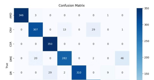
The system has been trained on a large and diverse OCT dataset, enabling it to detect subtle diagnostic features unique to each disease category. By recognizing these patterns automatically, the model supports early diagnosis and timely intervention for conditions that could otherwise lead to vision impairment or blindness.
This solution serves as a valuable clinical support tool, helping ophthalmologists improve diagnostic efficiency while maintaining high levels of accuracy.
Project Tips
This project provides an efficient and scalable solution for screening and diagnosing retinal diseases, helping eye care clinics optimize resources and improve patient outcomes.
- Accurate multi-class classification of eight retinal disease categories.
- Utilization of OCT imaging for non-invasive retinal diagnosis.
- Detection of subtle disease indicators such as fluid accumulation and retinal abnormalities.
- Reduced dependence on manual image interpretation during initial screening.
- Rapid diagnostic results that improve patient workflow management.
- Potential integration as an early-warning diagnostic tool in clinics and healthcare centers.
Overview & Challenge
The innovative aspect of this project lies in its ability to perform accurate multi-class classification across eight disease categories in addition to normal retinal conditions.
The model learns the normal anatomical structure of retinal layers and understands how different diseases alter those structures. By extracting spatial features from retinal layers and applying advanced deep learning algorithms, it can accurately classify the type of retinal abnormality present.
A key innovation is the system’s ability to handle the complexity of medical imaging data while delivering clear, actionable diagnostic results. The project represents a shift from descriptive diagnosis toward data-driven, measurable, and AI-assisted clinical decision-making.
Furthermore, it creates opportunities for closer collaboration between AI specialists and ophthalmologists, contributing to the future of intelligent healthcare systems.
Practical Implementation
The model can be used as a clinical decision-support tool for ophthalmologists by providing fast and accurate analysis of OCT scans. This facilitates early disease detection, accelerates treatment planning, and improves overall patient care outcomes.